
Fetal Medicine
Ultrasound scans
Viability scan
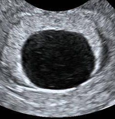
This is an ultrasound examination that is usually carried out vaginally at 6-10 weeks of pregnancy. The aims of this scan are to determine the number of embryos present and whether the pregnancy is progressing normally inside the uterus.This scan is useful for women who are experiencing pain or bleeding in the pregnancy and those who have had previous miscarriages or ectopic pregnancies.
The figure on the left shows a normal pregnancy at 7 weeks of gestation. The figure on the right shows the empty sac of an anembryonic pregnancy at 7 weeks.
Nuchal scan
This scan is carried out from 11 weeks to 13 weeks and six days. The scan is usually performed transabdominally but in a few cases it may be necessary to do the examination transvaginally.
Aims of the nuchal scan
- To date the pregnancy accurately. This is particularly relevant for women who cannot recall the date of their last period, have an irregular menstrual cycle, or who have conceived whilst breastfeeding or soon after stopping the pill. We measure the size of the fetus and from this we calculate the expected date of delivery.
- To diagnose multiple pregnancy. Approximately 2% of natural conceptions and 10% of assisted conceptions result in multiple pregnancy. Ultrasound scanning can determine if both babies are developing normally and if the babies share the same placenta which can lead to problems in the pregnancy. In such cases it would be advisable to monitor the pregnancy more closely.
- To diagnose major fetal abnormalities. Some major abnormalities may be visible at this gestation. However it will still be necessary to have a 20 week anomaly scan.
- To diagnose early miscarriage. Unfortunately, in 2% of women who attend for a nuchal scan it is found that the fetus has died, often several weeks before and without any warning. Couples will receive full counselling as to the possible causes of this problem and the options for subsequent measures that may be necessary.
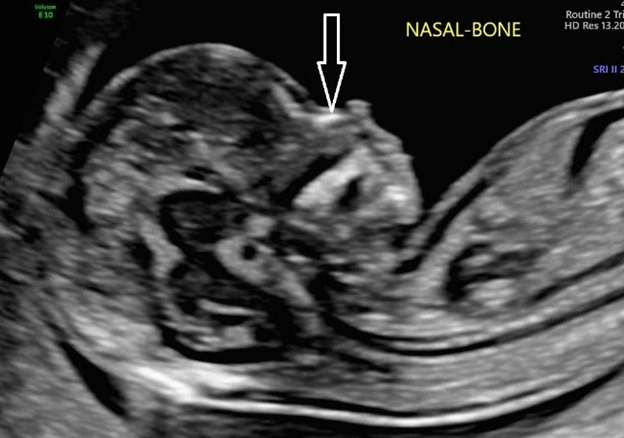
- To assess the risks of Down’s syndrome and other chromosomal abnormalities. Each woman will be given an estimate of her individual risk for this pregnancy. This is calculated by taking into account the age of the mother, measurement of two hormones in the mother’s blood and the scan findings of nuchal translucency thickness, nasal bone, blood flow through the fetal heart and ductus venosus and fetal abnormalities. Parents will receive full counselling concerning the significance of these risks and the various options for further investigations including invasive testing or the Harmony test.
Anomaly scan
This is a detailed scan at 20-24 weeks of pregnancy.
During the scan we examine each part of the fetal body, determine the position of the placenta, assess the amount of amniotic fluid, and measure fetal growth. Special attention is paid to the brain, face, spine, heart, stomach, bowel, kidneys and limbs. If any abnormalities are detected the significance of the findings will be discussed and the couple will be given the opportunity to have further counseling.
Cardiac scan
During the nuchal scan (11-13 weeks), the anomaly scan (20-24 weeks) and wellbeing scan (30-34 weeks) we routinely examine the fetal heart and connecting blood vessels.
A specialist examination of the fetal heart is recommended for:
- Women with family history of congenital heart abnormalities, those with diabetes mellitus and those taking antiepileptic drugs
- Fetuses with suspected heart defect and those with increased nuchal translucency or certain non-cardiac abnormalities detected during the routine scans
Cervical scan
This is a transvaginal scan to measure the length of the cervix.
It is recommended in women at high risk for preterm birth, including multiple pregnancies, those with a previous preterm birth, abnormalities of the uterus or previous cervical surgery. This examination is usually carried out at the time of the anomaly scan but in women with previous preterm birth it may be necessary to perform a series of scans starting from 16 weeks.
Wellbeing scan
This ultrasound scan is usually carried at about 32 weeks of pregnancy. Some obstetricians advise that this scan is offered to all women. Others reserve such scans for those women who have had previous complications of pregnancy such as pre-eclampsia, growth restriction, diabetes, stillbirth, and for those women who develop a problem during the course of their current pregnancy.
This scan aims to determine the growth and health of the fetus by:
- Measurement of the size of the fetal head, abdomen and thigh bone and calculation of an estimate of fetal weight
- Examination of the movements of the fetus
- Evaluation of the placental position and appearance
- Measurement of the amount of amniotic fluid
- Assessment of blood flow to the placenta and fetus by colour Doppler ultrasound
Invasive tests
Chorionic villus sampling
What is chorionic villus sampling?
- Chorionic villus sampling (CVS) involves the examination of chorionic villi (placental tissue). Both the baby and placenta (afterbirth) originate from the same cell and so the chromosomes present in the cells of the placenta are the same as those of the baby.
How is CVS done?
- Local anaesthetic is given. A fine needle is then passed through the mother’s abdomen and a sample of villi is taken. The needle is carefully observed using ultrasound scan.
- The procedure lasts 1 minute and afterwards we check that the fetal heart beat is normal.
What should I expect after the CVS?
- For the first couple of days you may experience some abdominal discomfort, period-like pain or a little bleeding. These are relatively common and in the vast majority of cases the pregnancy continues without any problems.
- You may find it helpful to take simple painkillers like paracetamol.
If there is a lot of pain or bleeding or if you develop a temperature please seek medical advice.
When can I expect to get the results?
- The results for Down’s syndrome and other major chromosomal defects are usually available within 3 days. The results for rare defects take 2 weeks. As soon as we get the results, we will call you to let you know.
Will the procedure need to be repeated?
- In approximately 1% of cases the invasive test will need to be repeated because the results are inconclusive.
What are the risks associated with the test?
- The risk of miscarriage due to CVS is about 1% and this is the same as the risk from amniocentesis at 16 weeks. If you were to miscarry due to the test, this would happen within the next five days.
- Some studies have shown that when CVS is performed before 10 weeks there is a small risk of abnormality in the baby’s fingers and/or toes. To avoid this risk we never perform CVS before 11 weeks.
Amniocentesis
What is amniocentesis?
- Amniocentesis involves the examination of cells in the fluid from around the fetus (amniotic fluid).
The cells in the amniotic fluid originate from the baby and so the chromosomes present in these cells are the same as those of the baby.
How is amniocentesis done?
- Amniocentesis involves passing a thin needle into the uterus in order to remove a small volume of amniotic fluid. The needle is carefully observed using ultrasound scan.
- The fluid is fetal urine and the amount removed by amniocentesis reaccumulates within a few hours.
- The procedure lasts 1 minute and afterwards we check that the fetal heart beat is normal.
What should I expect after amniocentesis?
- For the first couple of days you may experience some abdominal discomfort or period-like pain. You may find it helpful to take simple painkillers like paracetamol.
- If there is a lot of pain, bleeding, loss of fluid from your vagina or if you develop a temperature please seek medical advice.
When can I expect to get the results?
- The results for Down’s syndrome and other major chromosomal defects are usually available within 3 days. The results for rare defects take 2 weeks. As soon as we get the results, we will call you to let you know
What are the risks associated with amniocentesis?
- The risk of miscarriage due to amniocentesis is about 1% and this is the same as the risk from chorionic villus sampling. If you were to miscarry due to the test, this would happen within the next five days.
- Some studies have shown that when amniocentesis is performed before 16 weeks there is a small risk of the baby developing club feet. To avoid this risk we never perform amniocentesis before 16 weeks.
Recent Posts

March 29, 2019

March 29, 2019